PINPOINT
Endoscopic fluorescence imaging
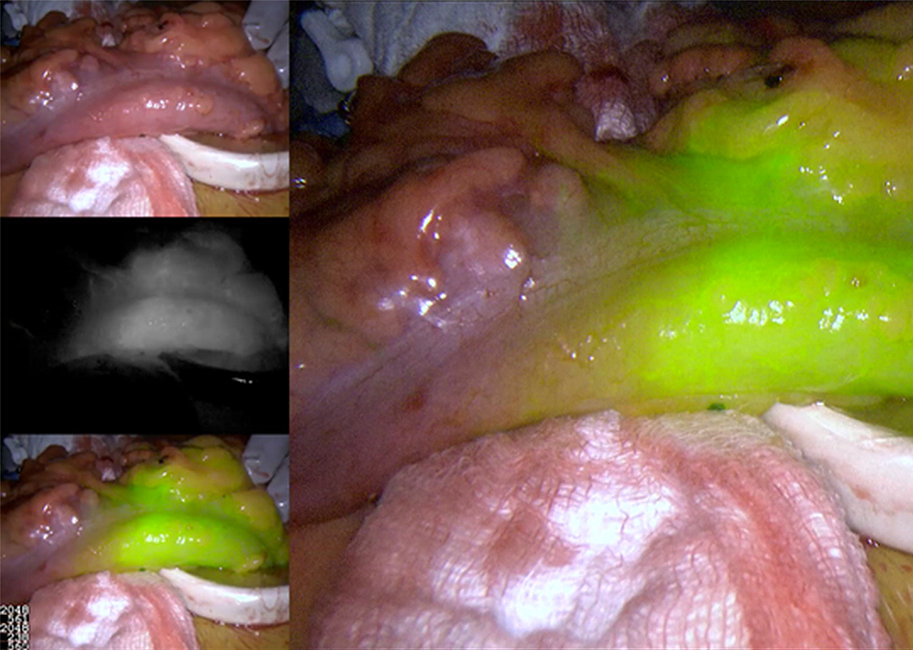
The PINPOINT Endoscopic Fluorescence Imaging System provides real-time high definition white light video and SPY Fluorescence Imaging during multiple surgical specialties.

Visualize tissue perfusion, biliary anatomy, and lymphatics in a different light.
PINPOINT is designed to offer simultaneous, real-time, high definition white light and fluorescence imaging through a single laparoscope. PINPOINT enables surgeons to perform routine visible light endoscopic procedures as well as further visually assess circulation including blood flow in vessels and microvessels, tissue and organ perfusion, and lymphatics and perfusion associated with tumors and tumor margins with near infrared fluorescence imaging during minimally invasive surgery.
The fluorescent imaging agent (indocyanine green) binds to proteins in blood, providing laparoscopic visualization of blood flow and tissue perfusion, and biliary anatomy.
* ICG is not provided by Stryker but is independently sourced by the customer. Customers should always consult the instructions for use of the manufacturer for specifications and use.
One technology,
multiple minimally invasive applications
multiple minimally invasive applications
PINPOINT offers high-definition, white light video with the added advantage of SPY Fluorescence Imaging technology, which has been demonstrated as beneficial in a variety of surgical applications.2, 3, 5
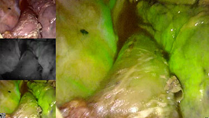
Colon resection
Assessing tissue perfusion may assist surgeons in making informed decisions that can positively impact outcomes.3
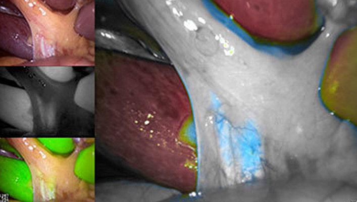
Laparoscopic cholecystectomy
Visualising biliary ducts and the critical view of safety, may be easier with PINPOINT.4
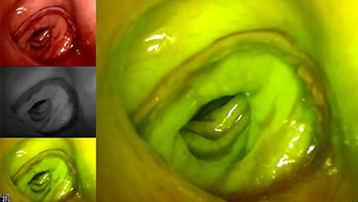
Minimally invasive esophagectomy
During esophagectomy, PINPOINT may help surgeons evaluate the gastric conduit.5
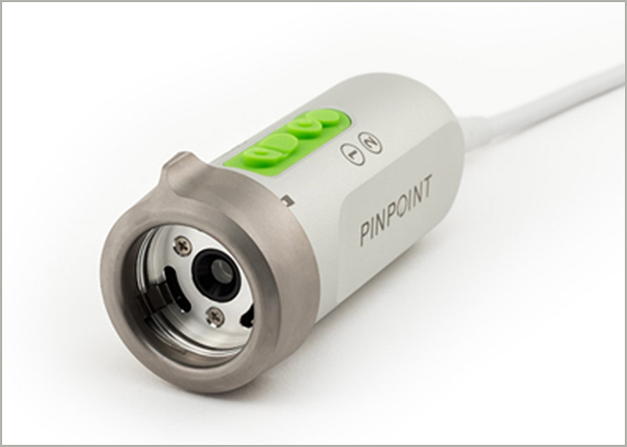
The PINPOINT camera
The camera features leading-edge sensor technology and its custom optics provide for vividly colored and high contrast images across all PINPOINT display modes.
Intraoperative perfusion assessment
Allows surgeons to visualize tissue perfusion and biliary ducts intraoperatively and in real-time.
Increased confidence
Fluorescence imaging may assist surgeons with critical decisions during surgery.3
Improved outcomes
Allows surgeons to assess perfusion, which may improve patient outcomes.3
Designed to
improving outcomes
improving outcomes
Published literature has long confirmed the significant social and economic burden associated with postoperative complications. More than 180 peer-reviewed medical journals have supported the use of SPY technology. Many of these studies have demonstrated an improvement in patient outcomes and a reduction in hospital costs as a result of SPY technology.2 For instance, the PILLAR II trial found that the use of SPY technology may assist in surgical decision making, which may reduce the occurrence of costly complications.3
References
1. Ott, Peter. Hepatic Elimination of Indocyanine Green with Special Reference to Distribution Kinetics and the Influence of Plasma Protein Binding. Pharmacol Toxicol. (1998)
2. Starker, Paul. “Using Outcomes Data to Justify Instituting New Technology: a Single Institution’s Experience.” Surgical Endoscopy, Dec. 2017, pp. 1586–1592.
3. Jafari MD, Wexner SD, Martz JE, McLemore EC, Margolin DA, Sherwinter, DA, Lee SW, Senagore AJ, Phelan MJ, Stamos MJ. Perfusion Assessment in Laparoscopic Left Sided/Anterior Resection (PILLAR II): A Multi-Institutional Study. JACS. Vol. 220, No. 1, January 2015.
4. Tsutsui, N. “Optimal Timing of Preoperative Indocyanine Green Administration for Fluorescent Cholangiography during Laparoscopic Cholecystectomy Using the PINPOINT® Endoscopic Fluorescence Imaging System.” Asian Journal of Endoscopic Surgery , Dec. 2017.
5. Fikfak, V. “Endoscopic Evaluation of Gastric Conduit Perfusion in Minimally Invasive Ivor Lewis Esophagectomy.” Int J Surg Case Rep, 2016, pp. 112–114.
SMACC 2020-26421