Resect and reconstruct in one surgery.
Discover PEEK Single Stage
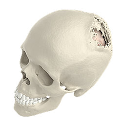
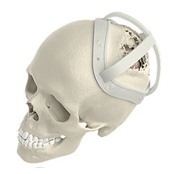
Plan ahead
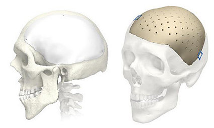
Through pre-surgical planning a predictive craniotomy is made. This leads to the design and manufacturing of a patient-specific implant and marking guides, which will be delivered to the hospital prior to surgery. Navigation files are also provided for every case.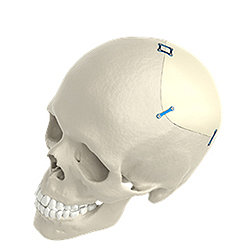
Individually designed. Personalised care.
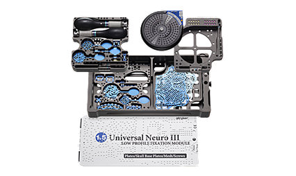
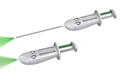
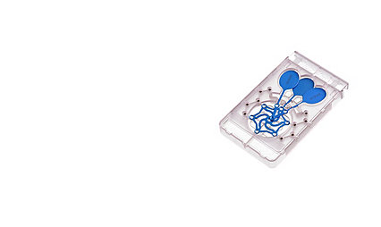
PEEK Single Stage offering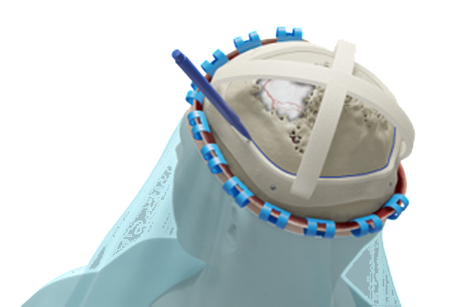
3D Systems marking guides
For PEEK Single Stage cases, 3D Systems offers surgical marking guides and anatomical models to assist with the craniotomy. Marking guides are available in either a cap (outer marking wall) or ring style (inner marking wall).
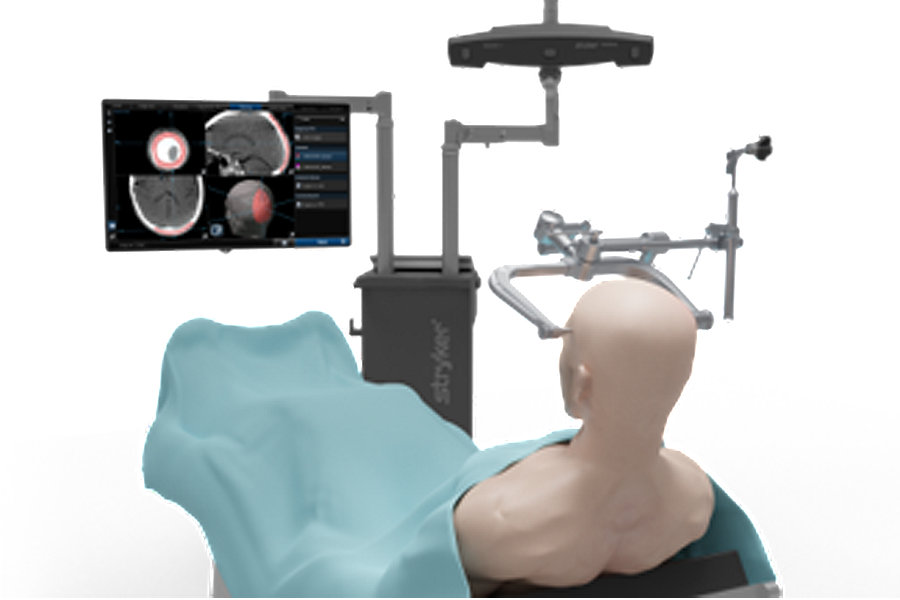
Navigation
Navigation can be integrated into PEEK Single Stage cases. Transfer the planned resection outline to the patient’s bony anatomy using compatible navigation system (STL and DICOM files).
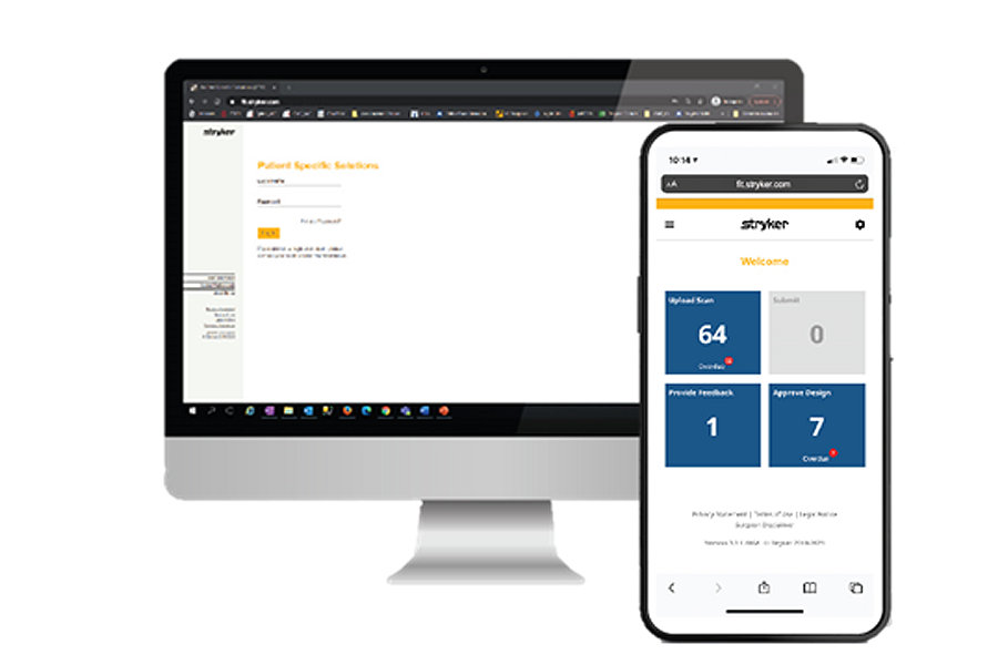
Easy workflow
PEEK Single Stage cases can be initiated and managed via desktop or mobile through our Stryker iD Portal. Each case is managed in four simple steps: 1. Case initiation, 2. Design session, 3. Case approval, 4. Shipment, which can all be tracked through the portal.
Clinically proven.
PEEK Single Stage clinical benefitsShorter operating time5
Single stage cranioplasty can avoid a second procedure and reduce overall operating time5. Longer operative times require prolonged anesthesia, which is known to be an independent risk factor for postoperative complications and should therefore be prevented7
Reduced burden on patient5
Since only one surgical procedure is required, hospitalisation time is reduced, and no helmet needs to be worn by the patient during revalidation to protect the underlying soft tissues. Also, immediate aesthetically satisfying reconstruction of the defect can be performed. Overall, single-stage cranioplasties therefore reduces the burden on the patient5
Functional & cosmetic outcomes2,3,4,6
The functional and cosmetic outcomes achieved with these surgeries are satisfying for surgeons and patients and remain stable throughout long-term follow-up2, 3, 4, 6.Instances most convincing to benefit from a single stage customised cranial implant may include nonhair-bearing regions, areas of actual or predicted male pattern hair loss, and potential areas of thin, irradiated scalps at risk for material extrusion3
Improved precision4
Complete resection of the tumor is the most important aspect to reach cure in patients with intraosseous meningiomas and can be planned preoperatively in a three-dimensional virtual model, in which also the implant is designed to ensure perfect fit to the resection line. Safety margins can be considered with this procedure to ensure resection of all tumorous tissue4
No image artifacts6,8,9
PEEK does not cause image artifacts and therefore allows imaging follow-up of the patients with either CT or MRI6. No negative consequences have been observed if radiotherapy is performed after insertion of PEEK implants8, 9
Get in touch today
Request your PEEK Single Stage brochure, clinical data, or schedule a product presentation.
Related products
- Jana Husse (2020): Clinical Evaluation Report on PEEK Cranial iD. Stryker Leibinger GmbH & Co KG.
- Alonso-Rodriguez E, Cebrián JL, Nieto MJ, Del Castillo JL, Hernández-Godoy J, Burgueño M. Polyetheretherketone custom-made implants for craniofacial defects: Report of 14 cases and review of the literature. J Craniomaxillofac Surg. 2015 Sep;43(7):1232-8. doi: 10.1016/j.jcms.2015.04.028. Epub 2015 May 8. PMID: 26032759.
- Berli JU, Thomaier L, Zhong S, Huang J, Quinones A, Lim M, Weingart J, Brem H, Gordon CR. Immediate Single-Stage Cranioplasty Following Calvarial Resection for Benign and Malignant Skull Neoplasms Using Customized Craniofacial Implants. J Craniofac Surg. 2015 Jul;26(5):1456-62. doi: 10.1097/SCS.0000000000001816. PMID: 26163837.
- Bianchi F, Signorelli F, Di Bonaventura R, Trevisi G, Pompucci A. One-stage frame-guided resection and reconstruction with PEEK custom-made prostheses for predominantly intraosseous meningiomas: technical notes and a case series. Neurosurg Rev. 2019 Sep;42(3):769-775. doi: 10.1007/s10143-019-01104-5. Epub 2019 May 4. PMID: 31055698.
- van de Vijfeijken SECM, Schreurs R, Dubois L, Becking AG; CranioSafe Group. The use of cranial resection templates with 3D virtual planning and PEEK patient-specific implants: A 3 year follow-up. J Craniomaxillofac Surg. 2019 Apr;47(4):542-547. doi: 10.1016/j.jcms.2018.07.012. Epub 2018 Jul 25. PMID: 30745010.
- Jalbert F, Boetto S, Nadon F, Lauwers F, Schmidt E, Lopez R. One-step primary reconstruction for complex craniofacial resection with PEEK custom-made implants. J Craniomaxillofac Surg. 2014 Mar;42(2):141-8. doi: 10.1016/j.jcms.2013.04.001. Epub 2013 May 18. PMID: 23688592.
- Phan K, Kim JS, Kim JH, Somani S, Di'Capua J, Dowdell JE, Cho SK. Anesthesia Duration as an Independent Risk Factor for Early Postoperative Complications in Adults Undergoing Elective ACDF. Global Spine J. 2017 Dec;7(8):727-734. doi: 10.1177/2192568217701105. Epub 2017 May 31. PMID: 29238635; PMCID: PMC5721997.
- Mangat NS, Kotecha A, Stirling AJ. The use of non-metallic implants to facilitate post-operative proton therapy in chondrosarcoma of spine. a case report. Orthopaedic Proceedings. 2012 94-B(Suppl. XXXVI).
- Sen CA. Our experiences and suggestions with PEEK marker use in prostate cancer radiotherapy. Contemp Oncol (Pozn). 2018;22(1):47-49. doi: 10.5114/wo.2018.74394. Epub 2018 Apr 3. PMID: 29692664; PMCID: PMC5909730.
2021-30453

